Products You May Like
The resolution of light microscopes has been given a huge boost thanks to the clever use of a common phenomenon in quantum physics.
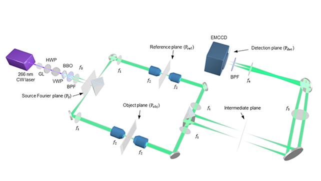
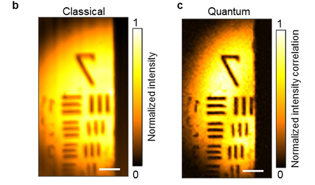
By sending entangled light down different paths and recombining their waves, it’s possible to peer at delicate objects more closely than ever before, effectively doubling their resolution without the usual complication of dramatically increasing the light’s energy.
It’s called quantum microscopy by coincidence (QMC), and was developed by researchers from California Institute of Technology (Caltech) in the US, who say that it’s particularly well suited to the examination of tissues and biomolecules to find disease or to study its spread.

“The combination of the improved speed, enhanced contrast-to-noise ratio, more robust stray light resistance, super resolution, and low-intensity illumination empowers QMC toward bioimaging,” the researchers write in their recently published paper.
Quantum entanglement describes correlations that exist between objects that have a shared history, prior to being observed. Just as two shoes bought at a store are correlated to fit a right foot and a left foot, particles can be mathematically correlated in a variety of ways too.
Only in a quantum system, things like shoes and electrons don’t truly settle on any of those states until they’re observed. They’re merely probabilities best described as a wave of maybes.
In QMC, the particles involved were photons, or light particles, which are known as biphotons once they’ve been entangled in a pair.
This was done through a special kind of crystal made from β-barium borate (BBO). When laser light is shone through the crystal, a very small fraction of the photons – only around one in a million – are converted into biphotons. The researchers were then able to separate the biphotons again, via a network of mirrors, lenses, and prisms.

One photon is sent through the material being studied, while the other photon is analyzed. Being entangled, the correlations measured in either photon will also say something about the journey of its partner. It’s the basis of another fairly novel technology called ghost imaging.
Yet this entangled dual-action has another trick up its sleeve. Biphotons have twice the momentum of photons, which also means their wavelengths are halved. Half the wavelength of light, in turn, means a higher resolution for the light microscope.
Ordinarily, light with shorter wavelengths also carries more energy, which at a certain point can damage the cells being studied. Think about the difference between harmless, long length radio waves, versus the more powerful, short length ultraviolet (UV) rays that can break DNA and cause sunburn.
In this case, while the entanglement process effectively halves the wavelength, it doesn’t increase the energy of the individual photons.
“Cells don’t like UV light,” says medical engineer Lihong Wang, from the California Institute of Technology (Caltech). “But if we can use 400-nanometer light to image the cell and achieve the effect of 200-nm light, which is UV, the cells will be happy, and we’re getting the resolution of UV.”
There’s room for improvement in this system too, including speeding up the imaging and being able to entangle more photons together, increasing the resolution even further. However, adding more photons means the likelihood of getting an entanglement – already one in a million – would go down even further.
Since entanglement is easily disrupted by interactions with the environment, increasing the number of photons in a system increases the likelihood that individual photons will interact with the environment rather than with one another.
While biphoton imaging has been tried before, the researchers behind the new setup have made several improvements throughout the process, and tested it out practically – making it one of the most promising techniques of its type.
“We developed what we believe is a rigorous theory as well as a faster and more accurate entanglement-measurement method,” says Wang. “We reached microscopic resolution and imaged cells.”
The research has been published in Nature Communications.
